 Co., Ltd")


Animal ultrasound machine, cart type animal ultrasound machine, pet store ultrasound machine, Jiangsu animal ultrasound machine manufacturer
Category:
Instrumentation/Non destructive testing instruments/Ultrasonic detector
Model:
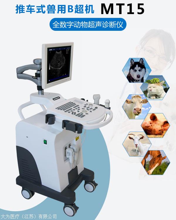
MT15
Brand:
Dawei Medical
Retail Price
120,000.00USD
重量
kg
- Product Description
-
Description :
Please discuss the product price directly by phone or in person.
Please discuss the product price directly by phone or in person.
Please discuss the product price directly by phone or in person.
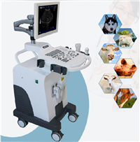
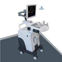
The MT15 animal ultrasound machine is a wheeled animal ultrasound machine with a 15 inch LED full view display. The pet ultrasound machine has stable performance, clear images, and supports ≥ 5 variable frequency probe frequencies. It can provide examination and diagnosis of liver, gallbladder, spleen, kidney, bladder, pregnancy and other tissues and organs for cattle, sheep, horses, pigs, dogs, cats, various poultry, experimental animals, small animals, and some aquatic animals.
1、 Introduction to the configuration of MT15 livestock ultrasound machine
Display: 15 inch LED full view display, freely adjustable display
● Probe frequency: Supports ≥ 5 types of frequency conversion, better reflecting clinical effects
Measurement software: provides measurement data such as obstetric gestational age, circumference/area, etc
● Probe interface: ≥ 2, dual interface and dual channel free switching
● Display magnification: Sixteen display modes; One click adjustment for various body types
● Host size: 740x520x1210mm
Net weight: 29.1kg Gross weight: 43.1kg
● Optional: 3.5MHz probe, 7.5MHz probe, micro convex probe, rectal probe
● Built in TF card and external USB storage, making image uploading more convenient
2、 Instructions for using MT15 livestock ultrasound machine
Ultrasound examination of abdominal organs:
Acute ultrasound examination of horses: Ultrasound examination of changes in acute symptoms of horses. Using a 3.5MHz convex array probe, the depth of the horse's abdomen is 20cm, with a depth focusing ability of 10-20cm. Abdominal ultrasound can be used to examine the symptoms of horse pain and correctly diagnose small intestinal stenosis obstruction.
Ultrasound examination of the anterior mesenteric artery in horses: examination of the anterior mesenteric artery and its branches through rectal ultrasound. Its imaging branches off from the aorta and can identify several branches.
Duodenal ultrasound examination of horses: Six healthy horses were fed with concentrated feed and hay, only hay, and fasted for 36 hours for ultrasound examination of the duodenum. Exploration location: Explore the intersection point between the line connecting the mouth to the nodule and the 16-17 intercostal space, and a duodenal image can be seen below the right kidney. Ultrasound shows a 5-layer structure: mucosal surface, mucosal layer, submucosal layer, muscular layer, and serosal layer.
Ultrasound examination of diaphragm rupture: Real time ultrasound is used to diagnose diaphragm rupture, and pleural effusion occurs after trauma. Ultrasound imaging shows gas filling the intestinal tract in the back and front of the diaphragm.
Ultrasonic examination of surgical incision on the lower midline of the horse's abdomen: Ultrasonic scanning is used to examine the healing of the incision on the lower midline of the horse's abdomen. Surgical incision examination 1-7 weeks after equestrian events. Use 5MHz and 7.5MHz probes for inspection. Visible incision drainage, edema, suture fistula formation, suture abscess, superficial opening (i.e. cracking), and incision hernia formation.
Ultrasound examination of bovine reticulum and small intestine: using a 3.5MHz linear array probe, located on the ventral side of the chest and between the sixth and seventh ribs. The examination includes the contour of the reticulum, contraction of the reticulum, and adjacent organs of the reticulum. Use a 3.5MHz linear array probe to explore the area from the hip nodule to the eighth intercostal space on the right abdominal wall; The transverse process of the spine reaches the white line (the white line indicates that the aponeurosis of both abdominal muscles combine along the midline of the abdominal floor to form a white line), and ultrasound can determine the movement of the small intestine and its contents.
Ultrasound examination of the liver and gallbladder system in cattle: ultrasound examination for extrahepatic bile stasis and cholangitis. Ultrasound examination of liver, gallbladder, severe bile stasis, intrahepatic bile ducts, cystic duct, and gallbladder with severe dilation. Measure the size, blood vessel diameter, and position of the cow's liver. Liver ultrasound is performed using a 3.50MHz linear array probe, located between the 12th, 11th, and 10th intercostal spaces on the right side. Measure the position and diameter of the dorsal and ventral edges of the liver, posterior vena cava, and portal vein at each intercostal space. Measure the ventral hepatic angle between the visceral and diaphragmatic surfaces, as well as the dorsal and peripheral edges of the gallbladder.
Calf umbilical cord ultrasound examination: Using a 5.0MHz or 7.5MHz probe, difficult to remove hernias and umbilical cord abscesses can be examined.
Skin ultrasound examination of cattle: Use a 7.5MHz probe to examine the skin of cattle. Measure the thickness of the nipple layer and reticular layer of the cow's skin to be 2mm and 4mm, respectively. When measuring skin thickness using ultrasound, pay attention to selecting the ultrasound contact medium and frequency.
Gastrointestinal ultrasound examination in dogs: Normal gastrointestinal ultrasound examination in dogs, with a gastric wall thickness range of 3mm to 5mm; The thickness range of the small and large intestine walls is 2mm to 3mm; The stomach and small intestine have regular peristalsis, while the colon cannot be seen. Ultrasound can identify the presence of the gastrointestinal wall layer, mucosal surface, mucosa, submucosal layer, and muscular layer, while the subserosal layer and serosal layer can sometimes be seen.
Ultrasound examination of the spleen in dogs: fine echogenic lines appear at the border of the spleen; The splenic parenchyma presents consistent speckled echoes, except for the anechoic cavity directly near the splenic hilum where blood vessels branch. There are fewer branches of splenic artery in the splenic hilum, while there are no arterial branches in the splenic parenchyma.
Exploration procedure: Dorsal transverse ultrasound imaging, with the ultrasound probe perpendicular to the abdominal wall between the left chest and ribs and its posterior abdomen. Longitudinal imaging is directed towards the ribs parallel to the lateral abdominal wall in the anterior direction.
Ultrasound examination of canine ascites and abdominal adhesions: After artificially inducing pneumoperitoneum, ultrasound examination of adhesions between the dog's stomach and intestines can distinguish a small amount of free ascites.
Ultrasound examination of canine adrenal gland: Ultrasound examination of the adrenal gland in healthy dogs, dogs without endocrine disorders, and dogs with adrenal hyperfunction. The experimental results showed that the length and width of the adrenal gland in normal dogs were 17.40mm × 4.10mm on the left and 16.70mm × 4.30mm on the right. In dogs without endocrine disorders (NED), the left adrenal gland was 19.00mm × 3.90mm on the left and 16.80 × 4.00mm on the right, with no difference compared to healthy dogs. The echo of the adrenal gland is weaker than the essential echo of the kidney.
3、 Application scope of MT15 livestock ultrasound machine
MT15 veterinary ultrasound machine is suitable for:
Cattle, sheep, horses, pigs, dogs, cats, various poultry, experimental animals, animals, and some aquatic animals
Application scope of MT15 veterinary ultrasound machine:
Examination and diagnosis of liver, gallbladder, spleen, kidney, bladder, uterus, pregnancy and other tissues and organs in small and medium-sized animals
AfterSalesService :
Key words:- Livestock B-ultrasound machine
More Products


