 Co., Ltd")


Small animal ultrasound detector, small animal endoscope, ultrasound, animal ultrasound, price, pet ultrasound machine manufacturer
Category:
Instrumentation/Non destructive testing instruments/Ultrasonic detector
Model:
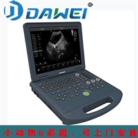
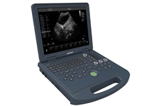
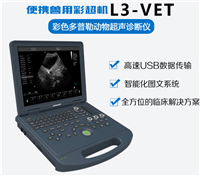
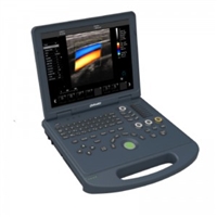
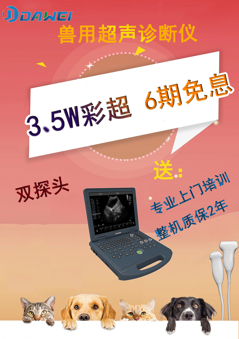
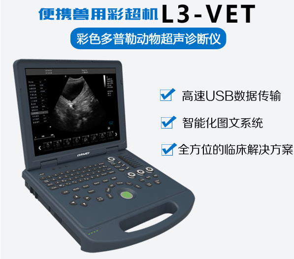
L3-VET
Brand:
Dawei Medical
Retail Price
35,000.00USD
重量
kg
- Product Description
-
Description :
Small animal ultrasound detector, small animal endoscopic ultrasound, animal B-ultrasound price, pet B-ultrasound machine, animal ultrasound diagnostic device is cheap, welcome to consult!
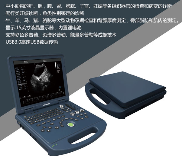
Introduction to Echo of Small Animal Ultrasonic Detector:
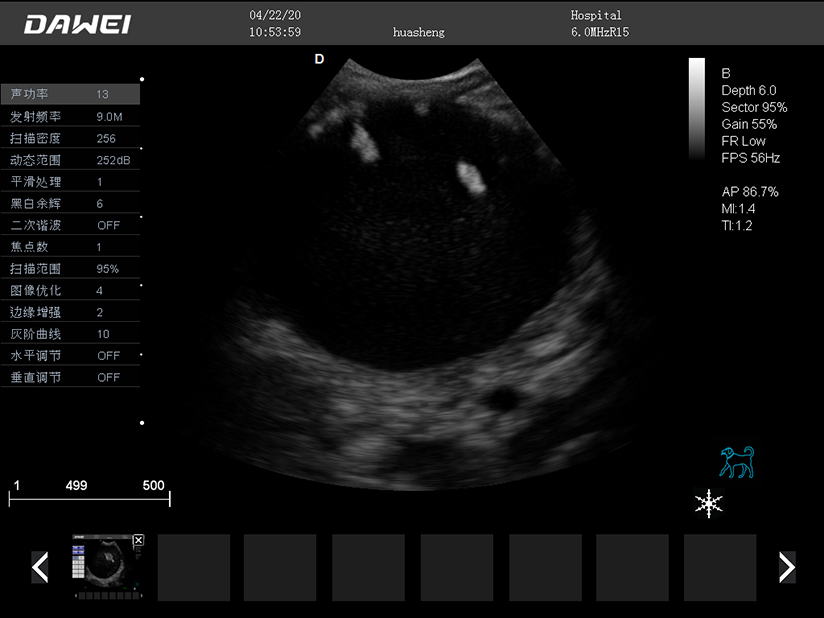
Echo intensity, echo size, echo density
The three parameters of echo - intensity, size, and density - describe the shape of the sound image, which can be divided into areas of no echo, low echo, and strong echo. Organizational characteristics are usually described in different ways.
Small animal ultrasound detectorNo echo
Organizations without acoustic interfaces, such as body fluids, bile, urine, blood, ingestion, and amniotic fluid, are all anechoic. They are displayed as black areas on the screen and printing paper.
Pathological fluid accumulation, such as ascites, pleural effusion, pericardial effusion, uterine effusion, or pus, abscess, hematoma, or cyst, without echo.
An area without echo is an echo free area. The received echo texture (point size) can be fine or rough, the intensity can be low or high, and the uniformity can be low or high.
Small animal ultrasound detectorhypoechoic
Structures with low echogenicity are characterized by subtle, scattered, weak or moderate to dark gray echoes.
Under normal circumstances, the renal medulla, intestinal muscles, and certain striated muscles are hypoechoic. It could also be low echo. Acute liver congestion or edematous pancreatitis may present with hypoechoic parenchymal structures
Small animal ultrasound detectorMid echo
Under normal circumstances, most parenchymal organs, such as the liver, spleen, pancreas, salivary glands, thyroid gland, adrenal gland, renal cortex, testes, and prostate gland, display moderate echogenicity. The sound and image of these organizations are composed of countless uniform, fine to semi rough, strong to moderate intensity, or bright to moderate brightness medium density echoes.
Animal ultrasound manufacturerIntroduction to High Echo
Tissue interfaces with significant differences in acoustic impedance can cause high-intensity reflections, which appear as white or bright gray images on screens or printed paper.
These structures are called strong echoes. The arterial wall, fibrous organ margin, interstitial fibrous tissue, and pericardium are usually highly echogenic. Calcified lesions, as well as the interface between bone tissue or gas and inflatable organs such as the trachea, esophagus, stomach, and small intestine, also exhibit high echogenicity. The total reflection of these interfaces can cause distant acoustic shadows.
Color Animal UltrasoundImaging image

AfterSalesService :
Key words:- Small animal ultrasound detector
More Products


